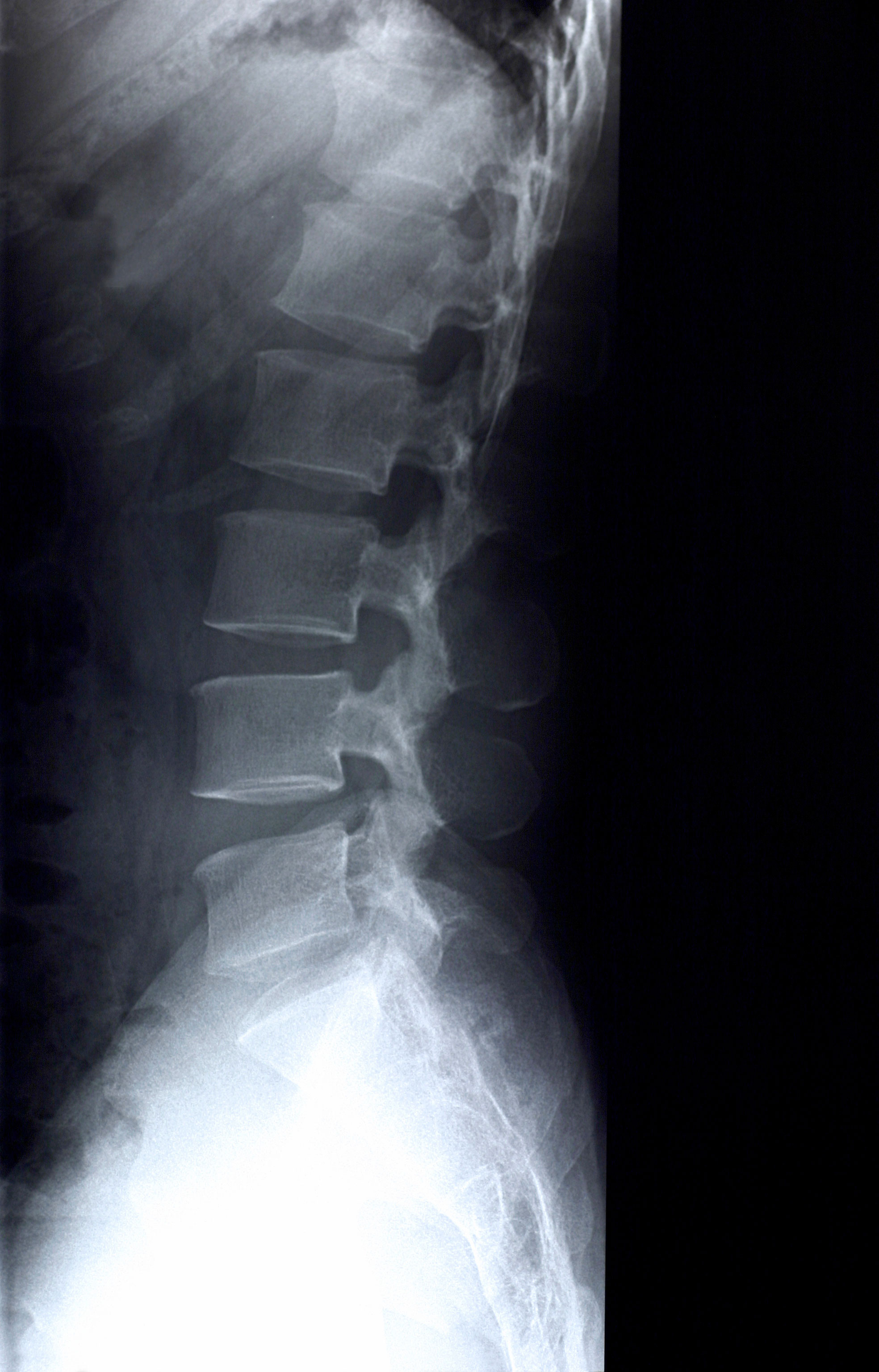

Magnetic resonance imaging (MRI): In addition to bones, an MRI is able to show soft tissues in the neck which aren’t visible in an X-ray or CT scan.

It may help your doctor detect even small changes in the bones and help see any calcifications. This allows your doctor to see the bones and discs in your neck. Computerized tomography scan (CT scan): This is a special kind of X-ray that uses a computer to provide cross-sectional images.An X-ray is often the step taken to see if additional imaging should be ordered. If your doctor is concerned you might have a fracture or some kind of bony deformity, especially if you had an accident or some kind of trauma, an X-ray may be ordered. X-ray: A neck X-ray, or cervical spine X-ray, uses radiation to get a flat picture of dense structures in your neck like bones and joints.When diagnosing neck pain, the three kinds of imaging you are most likely to encounter include: If your pain has persisted for years or if your doctor has any areas of concern after learning your history and conducting the physical exam, imaging studies may be ordered. Reflexes, strength, and sensation in different areas of your body will also be assessed. Your neck and back will be checked for any deformity, noting areas of tenderness or changes in range of motion.
#NORMAL NECK XRAY FULL#


 0 kommentar(er)
